Computer Vision in Medical Imaging (CVMI) Research
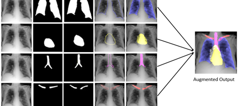
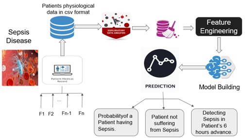
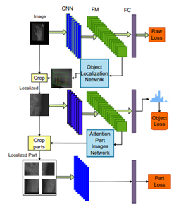
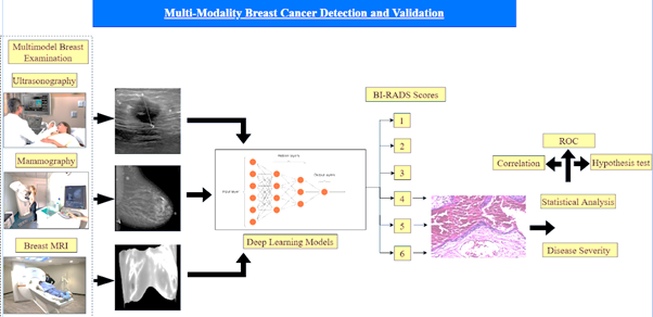
In “CVMI – Computer Vision in Medical Imaging” project, we wanted to make extensive use of computer vision in medical images (non-invasive imaging) to provide us better target lesion visualization, diagnosis, treatment and predication of various diseases. Computer vision can also help to find the texture, shape, contour and prior knowledge along with contextual information from medical image sequence and provide multidimensional information that benefits with better human understanding.
Powerful computer vision tools like image segmentation, visualization, registration, vision and machine learning, pattern classification, and keep track of targets lesions are much needed for quantitative analysis and study. Work done by a Radiologist often includes similar tasks i.e., finding a pattern of irregularity in bones, target tissues or tissues from images. This makes Computer Vision ideal choice for helping radiologist by automating some of the tasks, thus improving productivity. Our goals in this CVMI project is to automate the traditional tasks performed by clinician and radiologists such as lesion detection, segmentation, classification, and monitoring from clinical medical imaging using CV techniques in Indian context.

According to NASSCOM, by 2025 AI and data science will account for about 10 per cent of India’s GDP.
AI Specialists and Data Scientists fall under the highest-growth job roles of tomorrow according to LinkedIn’s 2020 Emerging Jobs Report
Adroit Market Research states that by 2025, data science will be a USD 178 bn industry.
Research Faculty
Dr. Sudipta Roy
Associate Professor, Artificial Intelligence & Data Science, Jio Institute, India
Researchers

Debojyoti Pal
Research Assistant, CVMI, Jio Institute

Mr. Komal Kumar
Research Assistant, CVMI, Jio Institute
Mr. Pankaj K. Jain
Research Associate, CVMI, Jio Institute

Mr. Snehasish Chakroborty
Research Assistant, CVMI, Jio Institute
Ms. Tanushree Meena
Research Assistant, CVMI, Jio Institute
Team Associates
Mr. Balakrishna Reddy
Vice President at Reliance AI-CoE
Dr. Kalyan Tadepalli
Student Mentor, Artificial Intelligence & Data Science Programme, Jio Institute | Consultant, AI Centre of Excellence, Reliance Jio, India
Highlights
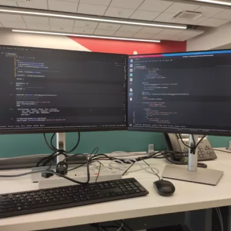
High performance computes for Research lab
A network of High Performance Computing services equipped with state-of-the-art computing and graphic processing power. Out system include Intel i9 processors, NVIDIA dedicated GPUs, and large RAM and storage for research purposes.
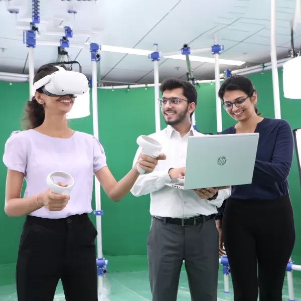
AR/VR Lab for research
A network of High Performance Computing services equipped with state-of-the-art computing and graphic processing power. Out system include Intel i9 processors, NVIDIA dedicated GPUs, and large RAM and storage for research purposes.
Know moreNews & Stories